Medical Testing
The Basis for Medical Treatment
The inner-ear mechanisms responsible for Dyslexia, LD or ADD and a score of related disorders and syndromes must first be demonstrated before medical treatment can be initiated. Most important, the pattern of diagnostic inner-ear-determined signs and symptoms characterizing each patient is essential for choosing the combination of medications most likely to be helpful.
SPECIFIC TESTS
- History
- Neurological Exam
- Electronystagmography
- Posturography
- 3-D Optical Scanning
- 3-D Auditory Scanning
- Audiological Testing
- Neuropsychological Exam
- Bender Gestalt Designs
- Figure Drawings
- Cerebellar Signs — Charts
-
Medical Testing — The Basis for Medical Treatment
The inner-ear mechanisms responsible for Dyslexia, LD, ADD and a score of related disorders and syndromes must first be demonstrated before medical treatment can be initiated. Most important, the pattern of diagnostic inner-ear-determined signs and symptoms characterizing each patient is essential for choosing the combination of medications most likely to be helpful.
Since only medical treatment is provided by The Levinson Medical Center for Learning Disabilities, only medical testing, especially of the inner-ear/cerebellar (CV) system, is performed — exceptions aside. (Educational and psychological testing are most helpful when tutoring is performed. Also, additional visual and related testing are provided to facilitate other therapeutic modalities and ensure diagnostic reliability). As a result, descriptions of the following non-invasive diagnostic tests and measurements are summarized to help you understand the primary procedures performed at this center:
History
The Self-Test: A Reliable Diagnostic Questionnaire
According to famed medical clinician Sir William Osler, history is 90% of diagnosis. Clearly, when over 35,000 dyslexics tell you what questions to ask, then history is truly a vital and effective diagnostic tool.
Take our Self-TestNeurological Testing
This clinical method consists of a series of standardized neurological tests commonly administered to assess the status of the integrated function of the inner-ear/cerebellar-vestibular (CV) system as well as other central nervous system (CNS) structures. Difficulties with any of the tests indicates a dysfunction within the CV or CNS systems.
The neurological mechanisms discovered by Dr. Levinson scrambling the Bender Gestalt and Goodenough drawing signals were found to be similar to those found diagnostic of cerebellar-vestibular (CV) impairment.
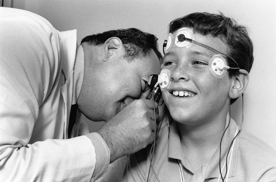
Electronystagmography (ENG)
The ENG is a standardized neurophysiological test in which eye movements are induced and measured under various testing conditions. Fine motor control and reflexive eye movements are controlled by the cerebellum and the inner-ear/vestibular system. As a result, the ENG can help determine whether or not an inner-ear/cerebellar abnormality exists.
Posturography
Posturography testing assesses overall balance function (sensory integration) as well as vision dependence, proprioception (internal senses) dependence, symmetry of weight bearing, lateral sway, and overall assessment of vestibular deficits. And its computer-generated scores also can serve as an objective measure of medication-triggered improvements. Also many of the symptoms of an inner-ear disorder (e.g., imbalance, dizziness, motion sickness, etc.) can sometimes result from other illnesses such as extreme stress and anxiety, dysfunction of cerebral and other related CNS structures. Posturography aids in this differentiation. A vestibular dysfunction produces a specific, quantifiable frequency and pattern of movement which is distinct from that caused by other disorders.
Neurological/ENG Test

A standardized neurological test.ENG/Posturography Testing
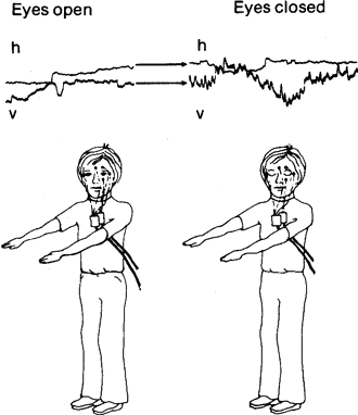
Recording abnormal eye (ENG) & balance movements due to inner ear dysfunction. Eyes-open and eyes-closed Romberg or balance positional ENG testing. The clinical insight that eyes-closed Romberg testing produced phobia-related and vestibular symptomatology led to the addition of a Romberg position ENG testing parameter. During eyes-closed Romberg balance testing, ENG was able to demonstrate the triggering of a vertical positional nystagmus. This evidence not only supported the clinical observations, but added a crucial new ENG testing parameter to the developing ENG methodology.
-
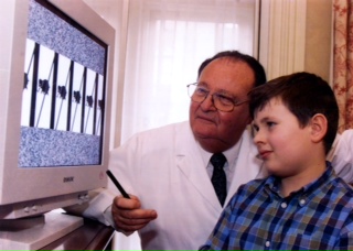
Optokinetic Testing — The 3-D Optical Scanner (refer to photo)
The inner-ear controls the eye’s ability to rapidly track and efficiently fixate targets. A dysfunction results in “clumsy" eye movements which can be objectively measured by the above noted ENG technique as well as a newly designed 3-D Optical Scanner. This new instrument is capable of rapidly and accurately determining "blurring speeds." Abnormal eye movements result in significantly reduced or pathological blurring scores in dyslexics as well as impaired fixation and movement illusions of stationary figures.
By utilizing medical instruments proven capable of diagnosing inner-ear/cerebellar dysfunction — including the 3-D Optical Scanner for detecting the eye tracking disorder characterizing dyslexics, it is possible to determine those with CV dysfunctioning and dyslexia in almost 100% of individuals tested, compensatory mechanisms aside.
How the 3-D Optical Scanner Works
When a sequence of elephants are accelerated across a computer screen, the eye is physiologically forced to track this sequence at a proportional speed so that clear vision can be maintained. Since inner-ear impaired dyslexics were found to have defective or clumsy eye movements, their eyes will not be able to “run” as fast as the elephants. As a result, they will blur-out the elephant sequence at speeds sharply reduced from those of normal children or adults, compensation aside. The blurring-speed is then a diagnostic indicator of an inner-ear/cerebellar dysfunction which predisposes children to dyslexia. Most important, this test is independent of the following typical variables complicating the usual “pencil and paper” tests, i.e., language, culture, socioeconomic status, prior tutoring, etc. Thus this test can be used to effectively screen young children before they begin to fail and before they begin to feel dumb, ugly, stupid, brainless, etc. Importantly, the 3-D Optical Scanner was found capable of improving tracking via conditioning techniques.
Audiological Testing
Audiological testing attempts to determine the presence or absence of middle ear and related hearing acuity problems. It evaluates (1) the pressure of the middle ear system, (2) the compliance or distensibility of the middle ear, (3) the acoustic reflexes of the ear drum, and (4) the ability of the patient to hear and distinguish different pitches at varying volumes.
3-D Auditory Rapid Scanning/Processing
The 3-D Auditory Scanner is a specifically designed instrument capable of measuring the speed at which accelerated phonetic sequences of words, phrases and sentences are blurred-out. This auditory blurring-speed is often significantly reduced in some dyslexics with inner-ear dysfunction. It thus serves as a diagnostic end-point. Important, Dr. Levinson also designed another method of compensating for this phonetic impairment and thus improving reading as well as auditory processing. Medical treatment can also significantly enhance this important function which coexists with auditory acuity.
By improving auditory scanning, it is possible to improve speech processing and clarity, irregardless of whether auditory acuity is normal or impaired.
3-D Optical Scanning Testing
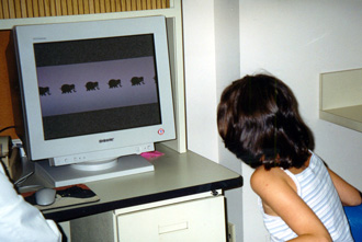
Observers report the speed at which accelerating elephants blur-out.
Observers report if/when a grid moving across stationary elephants trigger illusions of elephant-movement and/or elephant blurring. -
Neuropsychological Testing
Bender Gestalt & Figure Drawings
The copying of Bender-Gestalt and Goodenough designs significantly tests CV-determined and regulated visual-spatial processing and graphomotor abilities. These drawings highlight CV-based difficulties with tilting, articulation of related body parts, angle formation, rhythmicity and overall maturity of graphomotor coordination and output.
Click to enlarge
Figure 1. The Bender Gestalt stimulus cards. Children are requested to draw nine Bender Gestalt stimuli. For children older than ten years, the graphomotor drawing performance should closely approximate the illustrated Bender Gestalt stimulus figures. These figures thus served as the expected norm for children tested. Variations from this graphomotor norm suggest the presence of impaired dyslexic or CV drawing/writing mechanisms.
Click to enlarge
Figure 2. Bender Gestalt performance by a 10½-year-old dyslexic girl. Articulation and angle formation errors were triggered by cards A, 4, 7, & 8. Directional disturbances resulting in drifting, rotation, and angel formation errors were triggered by cards A, 1, 5, 7, and 8. In addition, card 6 triggered zig-zagging rather than a smooth rhythmic flow, and card 2 provoked graphomotor instability in maintaining constant size and space as a function of repetition and time.
Click to enlarge
Figure 3. Bender Gestalt performance by a 10-year-old dyslexic boy. Articulation and angle formation errors were triggered by cards A, 4, 7, and 8. Directional or drifting errors were triggered by cards 1 and 2. Card 6 provoked dysmetric or dyslexic zig-zagging rather than a smooth rhythmic flow.
Click to enlarge
Figure 4. Bender Gestalt and Goodenough figure drawings of a 10½-year-old dyslexic boy. Cards A, 4, and 8 were found to trigger angle-formation errors. Orientational and directional disturbances were provoked by card 2. Card 6 triggered zig-zagging. The figure drawing is simplified, detail is minimized and the arms are omitted in an effort to avoid the anxiety triggered by complex graphomotor tasks.

Click to enlarge
Figure 5 A–D. The somatically determined errors provoked when dyslexics are requested to draw a person. As noted, the figures are frequently tilted and details such as hands, fingers, feet and facial features are omitted. (D) A stick figure illustrates the simplification of the arms, legs, and trunk, although the facial features are detailed. These Goodenough figure drawing errors were viewed as indicative of underlying disturbances in CV-determined spatial, directional, graphomotor and proprioceptive integration. In addition, some of the simplification or omission errors were assumed to represent avoidance of inept performance and anxiety.CV Neurological Mechanisms and Signs
The neurological mechanisms discovered by Dr. Levinson to distort the Bender Gestalt & Goodenough drawing were found to be similar to those determining the diagnostic cerebellar-vestibular (CV) signs of impairment in the following tables:
Symptoms of Cerebellar Deficiency
Neurocerebellar Lesions Muscular hypotonia
Disturbances of posture
- Abnormal attitudes: Shoulder, body, and for head tilt or rotation
Static tremor
Disorder of movement (ataxia)
- Dysmetria
- Tremor (movement)
- Adiodokokineses
- The rebound phenomenon
- Associated movements
Ocular disturbances
- Weakness of conjugate ocular deviation
- Skew deviation
- Nystagmus
Disorders of articulation and phonation
Disorders of gait
Abnormalities of the reflexes
- "Pendular" knee-jerk
Barany's pointing test
Lesions of the Flocculonodular Lobe Balance and gait disturbances
- Vertigo
Data from Brain, 1955, pp. 52-55. The Most Common Symptoms of Cerebellar Deficiency
Division of the Cerebellum Responsible for Symptom Symptoms, in Order of Frequency Focculonodular Lobe Anterior Lobe of Corpus Cerebelli or Medial Part of Corpus Cerebelli Posterior Lobe of Corpus Cerebelli or Lateral Parts of Corpus Cerebelli 1. Disturbance in gait 2. Ataxia of isolated movements of upper extremity 3. Spontaneous nystagmus ? 4. Adiadochokinesis 5. Ataxia of lower extremity 6. Abnormal head posture 7. Hypotonia 8. Disturbance in station 9. Past-pointing and spontaneous deviation of the limbs ? 10. Stewart-Holmes phenomenon 11. Dysmetria 12. Tremor 13. Cerebellar speech disturbance 14. Pendular knee jerk 15. Cerebellar "fits" ? 16. Cerebellar catalepsy ? 17. Positive supporting reaction ? Classified according to the division of the cerebellum thought primarily responsible for the symptom when diseased or damaged (Dow and Moruzzi, 1958)